Brown Material on a Coral: Record the Signs First
Brown, stringy or gelatinous material can be worrying, especially beside changing tissue. Appearance alone does not establish a disease, and the priority is to describe what is happening clearly.
Record location and timing
Is the material emerging near a mouth, sitting on the surface, or beside an area where tissue is missing? Note when it appeared, whether it moves with the current and whether the affected area is expanding. Use a close photo plus a view of the entire coral.
Separate observations from explanations
Mucus, expelled material, deposits and deteriorating tissue are not interchangeable descriptions. A brown color or a familiar nickname cannot identify a pathogen. Avoid copying a medication or dip protocol from an image that only looks similar.
Gather context
- Record temperature, salinity and recent water tests.
- List changes in lighting, flow, additions and maintenance.
- Compare with older photographs.
- Avoid touching or scraping tissue to improve the view.
Escalate a rapid change
If tissue is visibly disappearing or several animals are declining, promptly contact an experienced aquarist or aquatic veterinarian with the images and timeline. This post supplies an observation checklist, not a treatment recipe.
Join the discussion
What observations did you make first?
Related Community reading
Sources and further reading
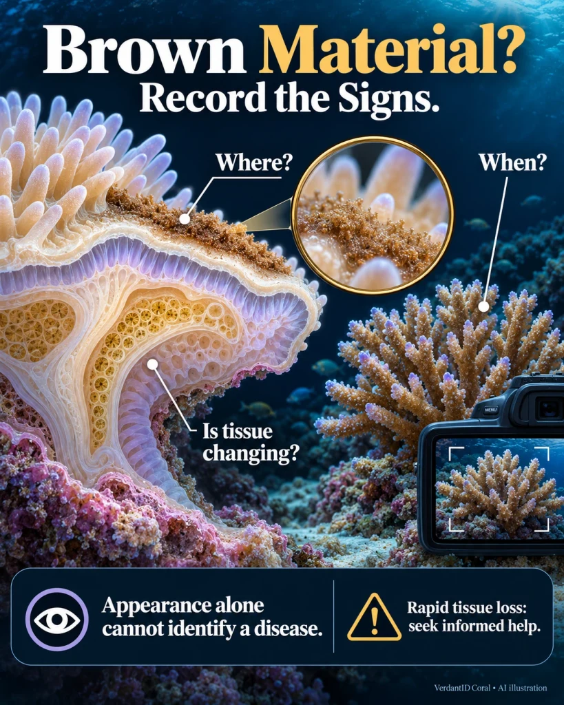
Source: Original Verdant guide