Duncan Coral Spotlight: Look Beyond the Open Polyp
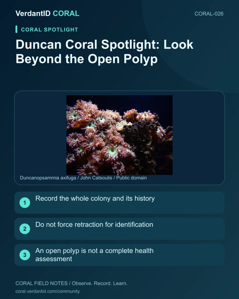
Duncan coral is commonly associated with Duncanopsammia axifuga. Its conspicuous polyps make it an appealing subject for regular photographs, but a single moment of extension is only one observation.
Notice the whole colony
An open polyp can obscure the branching skeleton beneath it. Photograph the colony's growth form when it is naturally visible; do not force retraction or expose tissue just to see the structure. Keep the supplier's identification and a reference source with the images.
Make feeding observations specific
If you record a response around feeding, include the food, approximate timing and the surrounding conditions. Retraction alone does not prove either successful feeding or a health problem. Avoid adding more food simply to reproduce an attractive response on camera.
Document a useful baseline
- Use the same angle and usual lighting for repeat images.
- Note changes in polyp number without disturbing the colony.
- Check that nearby tissue is not rubbing against rock.
- Keep measured readings and any placement changes with the record.
Keep care species-aware
A spotlight is an introduction, not a universal light, flow or feeding prescription. If the identification or a tissue change is uncertain, ask for help with clear photographs and history. The accompanying real photograph is identified as Duncan coral by its source.
Join the discussion
Do yours close when feeding?
Sources and further reading
Photo provenance: [John Catsoulis; public domain](https://commons.wikimedia.org/wiki/File:Duncanopsammia_axifuga_(whisker_coral).jpg).
Source: Original Verdant guide